Ultrasound of the Abdomen
- Onyait Reuben
- Jan 30, 2025
- 3 min read

Abdominal ultrasound is a non-invasive and painless medical imaging technique that uses sound waves to visualize the organs and structures within your abdomen. It's a valuable tool for diagnosing a wide range of medical conditions.
How it Works
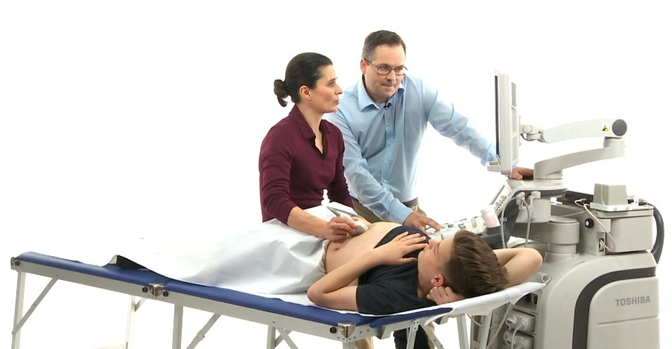
During an abdominal ultrasound, a Sonographer applies a gel to your skin and then moves a handheld device called a transducer over your abdomen. The transducer emits high-frequency sound waves that bounce off your internal organs, creating images on a screen.
What We Can See
Abdominal ultrasound allows us to examine various organs, including:
Liver: Assess size, shape, and detect abnormalities like tumors, cysts, or cirrhosis.
Gallbladder: Identify gallstones, inflammation (cholecystitis), or biliary duct blockages.
Pancreas: Visualize inflammation (pancreatitis), tumors, or cysts.
Spleen: Evaluate size and detect abnormalities associated with infections or blood disorders.
Kidneys: Assess size, structure, and identify kidney stones, hydronephrosis (swelling due to urine blockage), or tumors.
Aorta and Inferior vena cava (IVC): Visualize the major blood vessel in the abdomen and detect aneurysms (bulges) or dilatations.
Other structures: Ultrasound can also help visualize enlarged lymph nodes, fluid collections, abscesses, or masses within the abdomen.
Why Your Doctor Might Recommend an Abdominal Ultrasound
Abdominal ultrasound is a versatile tool used to investigate various symptoms and conditions, including:
Abdominal pain: To identify the cause of pain, such as gallstones, kidney stones, or appendicitis.
Abnormal blood tests: To further investigate abnormal liver function tests or other blood test results.
Palpable mass: To characterize a lump or mass felt during a physical examination.
Swelling or distention: To evaluate the cause of abdominal swelling or fluid buildup (ascites).
Fever: To look for sources of infection, such as abscesses or appendicitis.
Abdominal Trauma: To assess for internal injuries after an abdominal injury.
Suspected bowel obstruction: Used together with plain abdominal radiographs to identify dilatation, air-fluid and fluid-filled bowel loops.
Common Findings on Abdominal Ultrasound
Normal: The organs appear normal in size, shape, and structure.
Gallstones: Bright echoes within the gallbladder with acoustic shadowing.
Kidney stones: Bright echoes within the kidney with acoustic shadowing.
Liver cirrhosis: Changes in liver texture and size, with possible signs of portal hypertension.
Pancreatitis: Enlarged pancreas with altered texture and possible fluid collections.
Appendicitis: Enlarged appendix with possible signs of inflammation.
Aortic aneurysm: Widening of the abdominal aorta.
Prostate enlargement: The prostate volume measures high value than normal
Hemoperitoneum: Fluid (blood) in the peritoneum
Liver/spleen rupture: An haematoma with organ enlargement may be sign.
Renal trauma: Rapture of the renal capsule and associated haematoma.
Benefits of Abdominal Ultrasound
Non-invasive: No incisions or injections are required.
Painless: The procedure is generally painless.
Safe: No ionizing radiation is used.
Relatively inexpensive: Compared to other imaging techniques like CT scans or MRI.
Widely available: Ultrasound machines are available in most hospitals and clinics.
Preparing for Your Abdominal Ultrasound
You may be asked to fast for several hours before the exam, especially if the gallbladder is being examined.
Your Sonographer may ask you to drink plenty of water to fill your bladder, which can help visualize certain organs.
Wear loose-fitting clothing for easy access to your abdomen.
What to Expect During the Exam
You will lie on an examination table, and the technician will apply a gel to your abdomen.
The Sonographer/technician will move the transducer over your abdomen, applying gentle pressure.
You may feel some pressure or mild discomfort as the transducer is moved.
The exam usually takes about 30 minutes to an hour.
After the Exam
You can usually resume your normal activities immediately after the exam.
The radiologist will interpret the ultrasound images and send a report to your doctor.
Your doctor will discuss the results with you and determine if any further testing or treatment is needed.
In Conclusion
Abdominal ultrasound is a valuable diagnostic tool that can help your doctor evaluate your abdominal organs and diagnose a wide range of medical conditions. It is a safe, painless, and non-invasive procedure that can provide important information about your health. If you have any further questions or concerns, please do not hesitate to ask your Sonographer or doctor.


Comments