Ultrasound of Ambiguous Genitalia
- Onyait Reuben
- Feb 28, 2025
- 3 min read

A Case of Ambiguous Genitalia in a 28-Year-Old Adult: Insights from Ultrasound Imaging
Introduction
Ambiguous genitalia, a condition where the external genitalia do not have the typical appearance of either male or female, is often diagnosed in infancy. However, in rare cases, individuals may present later in life with subtle or previously unnoticed features. This blog post discusses a unique case of a 28-year-old adult who presented with ambiguous genitalia and the role of ultrasound in diagnosing the underlying anatomy.
Case Presentation
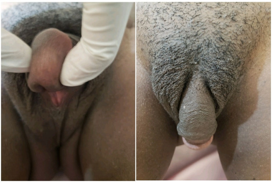
A 28-year-old individual presented to the clinic with concerns about a small penis and a vagina-like orifice beneath it, flanked by labia-like structures. The patient also reported palpable testes in the groin regions. Notably, there was no gynecomastia, and the patient was able to achieve erections and ejaculate. Vital signs were within normal limits: blood pressure was 115/73 mmHg, pulse rate was 48 bpm, and oxygen saturation was 99% in room air.
Clinical Findings and Ultrasound Imaging
Given the atypical presentation, the physician requested an ultrasound to evaluate the internal anatomy. The ultrasound findings were as follows:
Testes: Bilateral testes were identified in the supra-scrotal region. The testes exhibited a multitude of echoes, suggesting normal testicular tissue with no evidence of malignancy or structural abnormalities.
Uterus and Cervix: A small uterus and cervix were visualized posterior to the urinary bladder. The cervical canal was seen communicating with the vaginal orifice located at the base of the penile shaft.
Adnexal Regions: No ovarian tissue was identified in the adnexal regions, ruling out the presence of functional ovarian structures.
External Genitalia: The ultrasound confirmed the presence of a small penis with a vaginal orifice and labia-like structures, consistent with the external findings.
Ultrasound Images


Ultrasound Scan Report
Findings:
Bilateral testes identified in the supra-scrotal region with normal echotexture.
Small uterus and cervix visualized posterior to the urinary bladder.
Cervical canal communicates with the vaginal orifice at the base of the penile shaft.
No ovarian tissue identified in the adnexal regions.
Conclusions:
The ultrasound findings are consistent with a diagnosis of 46,XY disorder of sexual development (DSD), specifically persistent Müllerian duct syndrome (PMDS). This condition is characterized by the presence of Müllerian duct structures (uterus, cervix, and upper vagina) in an individual with a 46,XY karyotype and otherwise male external genitalia. The presence of bilateral testes and the absence of ovarian tissue further support this diagnosis.
Discussion
Persistent Müllerian duct syndrome is a rare form of DSD caused by mutations in the anti-Müllerian hormone (AMH) gene or its receptor. Individuals with PMDS typically have normal male external genitalia but retain internal Müllerian structures due to the failure of AMH signaling during fetal development. In this case, the presence of a small penis, vaginal orifice, and labia-like structures suggests a partial masculinization of the external genitalia, which is atypical for PMDS and may indicate a variant presentation.
The ability to achieve erections and ejaculate suggests functional testes and adequate testosterone production. However, the absence of gynecomastia indicates normal estrogen levels, which is consistent with the lack of ovarian tissue.
Management and Follow-Up
The patient was referred to a multidisciplinary team, including endocrinologists, urologists, and geneticists, for further evaluation and management. Hormonal assays, karyotyping, and genetic testing were recommended to confirm the diagnosis and guide treatment. Psychological support was also provided to address any concerns related to gender identity and sexual function.
Conclusion
This case highlights the importance of ultrasound imaging in diagnosing complex cases of ambiguous genitalia. The ability to visualize internal structures such as the uterus, cervix, and testes provides critical information for accurate diagnosis and management. Early and precise diagnosis is essential for guiding treatment, addressing psychological concerns, and improving the quality of life for individuals with DSD.
Key Takeaways
Ambiguous genitalia can present in adulthood, requiring a thorough diagnostic workup.
Ultrasound is a valuable tool for visualizing internal reproductive structures and guiding diagnosis.
Persistent Müllerian duct syndrome is a rare but important consideration in 46,XY individuals with retained Müllerian structures.
Multidisciplinary care is essential for managing complex cases of DSD.
By sharing this case, we hope to raise awareness about the diagnostic challenges and management strategies for ambiguous genitalia, ultimately improving patient outcomes.


Comments