Ultrasound Diagnosis of Anencephaly at 20 Weeks in a 28-Year-Old G2P1+0 Patient
- Onyait Reuben
- Jun 13
- 3 min read

A Case Study


Introduction
Anencephaly is a severe neural tube defect (NTD) resulting from failure of the anterior neuropore to close during embryonic development. It is characterized by the absence of the cranial vault (calvarium) and cerebral hemispheres, leading to nonviability. Early diagnosis via prenatal ultrasound is crucial for timely counselling and management. This blog presents a detailed sonographic case of anencephaly detected during a routine fetal anatomical scan at 20 weeks of gestation.
Clinical Presentation
A 28-year-old woman, gravida 2 para 1+0, presented to the antenatal clinic for her second antenatal visit at approximately 20 weeks of amenorrhea (WOA). She reported perceiving fetal movements and had no complaints of pain or bleeding.
General Physical Examination Findings:
General condition: Good
Vital signs:
Blood Pressure: 115/83 mmHg
Pulse: 97 bpm
Respiratory Rate: 18 bpm
SpO₂: 98% on room air
Abdominal Examination:
Abdomen distended and moving with respiration
Fundal height: 20/40
Non-tender, soft, no organomegaly
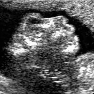
Ultrasound Findings
Obstetric Ultrasound Scan at ~20 Weeks Gestation
Fetal number and lie: Single live intrauterine fetus in oblique presentation
Cranial findings:
Absent calvarium (cranial vault)
Exposed disorganized brain tissue (exencephaly)
Classic "frog-eye" facial appearance due to absent skull bones and protruding orbits
Fetal heart rate: 156 bpm
Estimated fetal weight (EFW): 381.4g (± 61.03g)
Gestational age estimation: 20 weeks 5 days (AGA – appropriate for gestational age)
Amniotic fluid index: Adequate, Deepest Vertical Pocket (DVP): 7.56 cm
Placental location: Fundal, intact and normally attached
No other structural anomalies identified in spine, abdomen, limbs, or thorax
Ultrasound Report Summary
Patient: [Redacted]
Age: 28 years
Gravida/Parity: G2P1+0
Gestational Age: 20W5D (by ultrasound)
Indication: Routine fetal anatomical survey
Scan Findings:
Single live intrauterine fetus with:
Absent cranial vault (anencephaly)
Exposed brain tissue
Characteristic facial features consistent with "frog-eye" appearance
No other gross anomalies noted
Adequate amniotic fluid
Fundal placenta, intact
Fetal biometry consistent with 20W5D gestation
Fetal heart activity present at 156 bpm
Estimated fetal weight: 381.4g ± 61.03g
Conclusion:
Ultrasound findings are consistent with anencephaly, a lethal neural tube defect.
The anomaly is incompatible with extrauterine life.
Multidisciplinary counselling and appropriate management options are recommended.
Laboratory Findings
Blood Group: A+
Complete Blood Count (CBC):
Hemoglobin: 10.4 g/dL
White Cell Count: 3.8 x10⁹/L
Platelets: 113 x10⁹/L
Coagulation Profile:
Bleeding time: 2 minutes 34 seconds
Clotting time: 7 minutes 45 seconds
Management Plan
Given the confirmed diagnosis of anencephaly and the non-viability of the fetus, the following multidisciplinary management steps were taken:
Counselling: The patient and her partner were sensitively counselled on the diagnosis, prognosis, and available options. Emotional support and grief management were initiated.
Admission: The patient was admitted to the Gynaecology ward for termination of pregnancy.
Medical Termination:
Cervical ripening with Dinoprostone 2 mg
Antibiotic prophylaxis:
Flucloxacillin 500 mg TDS × 5 days
Metronidazole 400 mg TDS × 5 days
Post-Abortion Care Planning: Follow-up arranged with obstetric team and psychological support services. Neural tube defect prevention strategies and folic acid supplementation were discussed for future pregnancies.
Discussion and Insight
Anencephaly can be detected as early as the first trimester through transvaginal sonography; however, this case highlights the importance of the routine fetal anatomical survey at 18–22 weeks, especially for patients who may not have had early scans.
The hallmark features absence of calvarium, disorganized brain matter, and frog-like face are classic sonographic indicators. While the fetus was alive at the time of scanning, the condition is universally fatal. Prenatal identification allows for early and humane termination options, while also offering the opportunity to address maternal care and future pregnancy planning.
Conclusion
This case emphasizes the crucial role of mid-trimester fetal anomaly scanning in detecting lethal congenital anomalies such as anencephaly. Ultrasound professionals play a key role in early detection, sensitive communication, and coordinated care delivery. Continued advocacy for timely prenatal visits and first-trimester scans remains essential in improving maternal-fetal outcomes.
If you're a student or practitioner looking to improve your understanding of fetal neurosonography, especially in identifying neural tube defects, stay tuned for upcoming case-based tutorials and scan review sessions.


Comments