Ambiguous Genitalia Ultrasound case Report
- Onyait Reuben
- Feb 28, 2025
- 2 min read

Updated: Apr 29, 2025
Ultrasound Case Report: Ambiguous Genitalia in a 28-Year-Old Young Adult
Introduction
Ambiguous genitalia is a rare condition that presents diagnostic and clinical challenges. Ultrasound plays a crucial role in assessing internal reproductive structures, guiding further evaluation and management. This case details the ultrasound findings of a 28-year-old male who presented with a small penis, a vaginal-like orifice beneath it, and labia-like structures on either side.

Clinical Presentation
A 28-year-old male presented to the clinic with concerns regarding genital anatomy. Physical examination revealed:
Small penis with a vaginal-like orifice at the base
Labia-like structures bilaterally
Palpable testes in the groin regions
No gynecomastia
Normal sexual function, with the ability to achieve erection and ejaculation
Vital signs: BP 115/73 mmHg, PR 48 bpm, SpO2 99% on room air
Ultrasound Findings
A comprehensive ultrasound examination was performed, revealing the following findings:
Testes: Bilateral testes were identified in the supra-scrotal regions, exhibiting multiple internal echoes, suggesting possible microlithiasis or tubular ectasia.
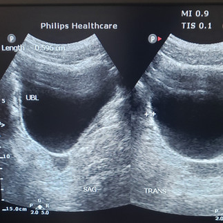
Uterus and Cervix: A small uterus with a defined cervix was visualized posterior to the urinary bladder.
Vaginal Canal: The cervical canal was seen extending and communicating with the vaginal-like orifice located at the base of the penile shaft.
Ovaries: No ovarian tissue was identified in the adnexal regions.
Other Findings: The urinary bladder appeared normal, with no structural abnormalities or mass lesions.
Ultrasound Images


Ultrasound Report
Patient Information:
Name: [Redacted]
Age: 28 years
Clinical Indication: Evaluation of ambiguous genitalia
Findings:
Bilateral testes are present in the supra-scrotal regions with multiple internal echoes.
A small uterus and cervix are visualized posterior to the urinary bladder.
The cervical canal communicates with the vaginal-like orifice at the base of the penile shaft.
No ovarian tissue is identified in the adnexal regions.
The urinary bladder appears normal.
Conclusion:
Imaging findings are consistent with disorder of sexual development (DSD), possibly persistent Müllerian duct syndrome (PMDS).
Further evaluation with karyotyping, hormonal assays, and genetic counseling is recommended for definitive diagnosis and management planning.
Discussion
Persistent Müllerian duct syndrome (PMDS) is a rare form of DSD in which males with 46,XY karyotype have retained Müllerian structures (uterus and fallopian tubes). This condition often presents with cryptorchidism or inguinal hernias, and in some cases, ambiguous genitalia.
In this patient, the presence of bilateral testes and a small uterus suggests PMDS. The absence of ovarian tissue and the presence of a vaginal-like orifice communicating with the cervix further support this diagnosis. The patient’s ability to achieve erection and ejaculation indicates functional male reproductive anatomy despite the presence of Müllerian structures.
Recommendations
Endocrine Assessment: Hormonal profile including testosterone, anti-Müllerian hormone (AMH), and luteinizing hormone (LH) levels.
Genetic Testing: Karyotype analysis (likely 46,XY) and evaluation for specific genetic mutations associated with PMDS.
MRI Pelvis: To further delineate the anatomy of internal structures.
Surgical Consideration: Consultation with a multidisciplinary team (urology, endocrinology, and genetics) to determine potential surgical management, including orchidopexy or Müllerian structure removal.
Psychosocial Support: Counseling and education regarding the condition and potential future interventions.
Conclusion
This case highlights the importance of ultrasound in evaluating ambiguous genitalia and disorders of sexual development. A multidisciplinary approach is essential for accurate diagnosis, appropriate management, and optimal patient outcomes.


Comments